
Our services
Fluoroscopy
Fluoroscopy is a technique that shows a continuous x-ray of the body like an x-ray movie, enabling the radiologist to view x-rays in real time.
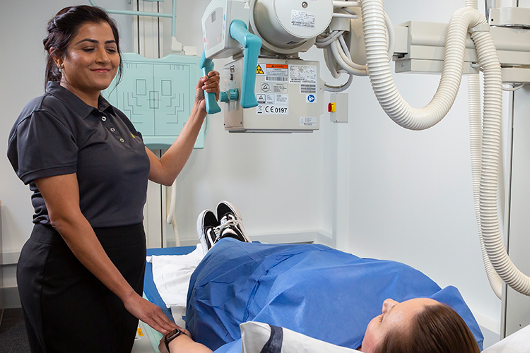
What is Fluoroscopy?
Fluoroscopy is a technique that shows a continuous x-ray of the body like an x-ray movie, enabling the radiologist to view x-rays in real time.
Fluoroscopy enables radiologists to assess many body systems including bones, joints, lungs, the digestive and urinary tracts, and the reproductive system.
Contrast media, such as barium sulphate and iodine-based dyes, are used to fill the area of interest, or outline soft tissue organs for more detailed visualisation and improved assessment.
Types of Fluoroscopy
Arthrography assesses bony joints such as; the hip, shoulder, ankle or knee. Any excess joint fluid might be withdrawn with a needle before contrast media is injected. After the contrast is injected, the patient might be asked to move the joint around to evenly distribute the contrast within the joint.
Injecting contrast media and/or air into a joint space enables the radiologist to assess the cartilage, ligaments, tendons and other structures. Once the contrast has been introduced into the joint more detailed images are acquired using fluoroscopy, CT or MRI.
Patients will be instructed to cease any blood thinners, and arrange transportation to and from the appointment.
Barium studies enable the radiologist to assess the oesophagus, upper gastrointestinal tract and stomach and as the contrast (Barium) shows on X-ray.
Barium is a chalky mixture of which is swallowed by the patient. As this mixture slowly travels through the intestinal lining, X-rays are taken to allow the radiologist to identify areas of abnormality such as ulcers, inflammation or tumours.
Generally you will be required to fast for 4 hours prior to the appointment so food does not obstruct the view.
Cystogram is used to assess the outline of the bladder in patients who are suspected of having urinary reflux, which means urine in the bladder is passed back through the ureters in the wrong direction.
Contrast media that is visible on X-ray is introduced through a small catheter to outline the bladder. Most patients do not find the procedure painful, however my experience some discomfort during this process.
No special preparation is required for this exam, and you can return to usual daily activities straight after.
Hysterosalpingography is used to assess the female reproductive system in women with infertility. Contrast medium is injected into the uterus through a small catheter. The passage of contrast through the uterus and fallopian tubes is followed using fluoroscopy. The test will show the shape of the intrauterine cavity, the width of the fallopian tubes and any blockage or flow restriction.
There might be some discomfort as the fluid passes through the tubes. Sometimes, the passage of the fluid through the tubes is enough to clear debris and allow a pregnancy to occur.
HSG’s are best performed one (1) week after menstrual cycle but before ovulation.
About Your Test
Before your appointment
The preparation prior to the scan is dependant upon the procedure required, area of interest and your medical history.
This may include fasting (generally 4 hours), however this will be explained to you at the time of the booking.
Generally, you will be asked to remove any jewellery, or clothing with metal clips, zips or buttons as these show up on x-rays.
If you need to remove any clothing you will be given a gown to wear.
On the day
Depending on which test your doctor has requested a contrast agent may be given which is introduced through ingestion, an enema, or an injection through a vein, usually at the elbow.
Our staff will explain the procedure to you.
You will be asked to lie on an x-ray table in a position that enables optimal visualisation of the area of interest, and you may be asked to hold your breath for a few seconds, to reduce movement.
The length of the examination varies according to the type of type of procedure and the body part being examined, but you should expect to be with us for at least an hour.
After your appointment
Any post-procedure care will be fully explained at the time of booking.
For many studies you can return to normal activities straight away, however, for some you may be required to arrange transport to and from your appointment.
You should contact your doctor if you notice any pain, redness, or swelling at the injection site as this could be an allergic reaction.
One of our radiologists will interpret the scan and provide your referring doctor with a comprehensive report about the findings.
The results of your scan will be sent directly to your referring doctor, and it is very important you book a timely follow-up appointment to discuss your results.
Frequently Asked Questions
Preparation will depend on the type of procedure, but our friendly booking staff will advise you when you book your appointment. Please be sure to bring along any previous x-rays, CT scans, ultrasound or MRI examinations which you may have available. They provide valuable additional information to the attending Radiologist performing your procedure. All imaging will be returned to you.
At the time of the procedure on the day, our healthcare team will discuss the detail of what will happen during the procedure.
Prior to your scan and to ensure optimal imaging, you may be asked to change into a gown. A change cubicle will be provided to ensure your privacy and you will be asked to bring your belongings will you, carry baskets are provided. You will be asked to place your belongings in a suitable location within the room for the duration of your scan. After your scan, you will be provided with a change cubicle to ensure your privacy. Please ensure you have all your belongings with you prior to leaving the department. If you accidentally leave anything behind, please contact our staff to advise and we will endeavour to locate your belongings and return them to you.
Each procedure is different and the time may vary depending on complexity.
Our friendly reception staff will advise you on the cost of your procedure at the time of your booking. If your exam is Medicare eligible, you will be bulk billed.
Patient Safety
Because of the nature of this procedure, they are only performed our 2 hospital sites.
Any risks involved with the procedure will be explained at the time of booking, and by our nurses on the day.
As Fluoroscopy uses X-ray, our staff are highly trained to use the lowest radiation dose possible for high quality images, minimalizing radiation exposure and decreasing risks involved.
The radiographer will only scan the area/s of concern, avoiding unnecessary exposure. The benefits of a fluoroscopy outweigh the risks involved.
Pregnant patients should avoid having a fluoroscopy in order to avoid exposing the fetus to unnecessary radiation.
The results of your scan will be sent directly to your referring doctor, and it is very important you book a timely follow-up appointment to discuss your results.