
Our services
Nuclear Medicine
Nuclear Medicine is a branch of diagnostic imaging that uses radio-pharmaceuticals to map the function of many organs and body systems. Common areas are the heart, bones, lungs, kidneys, liver, stomach and thyroid gland.
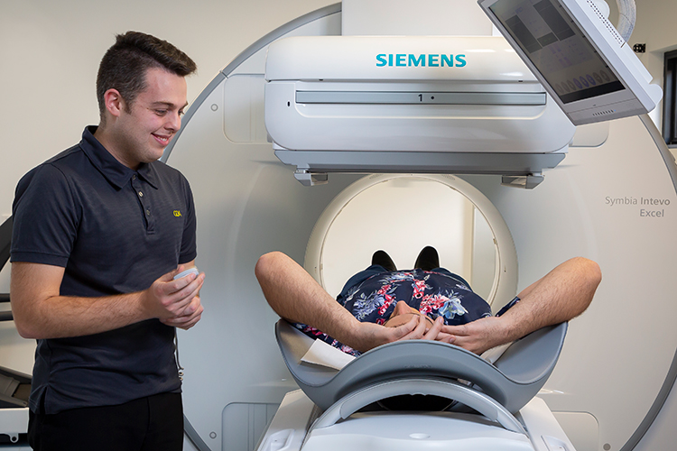
What is Nuclear Medicine?
The tracer, is usually injected into a vein and concentrates in the area of interest. A gamma camera moves closely over the area, building an image from the radiation emitted. A complex digital system then analyses, stores and displays the images that are evaluated by our medical imaging specialist doctors.
Nuclear Medicine can provide a rapid and accurate diagnosis of a wide range of medical conditions when used alone or in conjunction with other imaging tests.
All Apex Radiology nuclear medicine technologists are accredited and registered with Australian Health Practitioner Regulation Agency (AHPRA).
Types of Nuclear Medicine Tests
Dependent upon the request from your doctor, the procedure involves the injection, swallowing or inhalation of a small amount of a chemical substance tagged with a radioactive tracer – a radiopharmaceutical.
The radiopharmaceuticals give off gamma rays and are designed to concentrate in the part of the body being investigated, where there could be an abnormality or disease, e.g. skeleton, lungs, heart or liver.
A special camera – a gamma camera then moves around the body, providing a series of images of the radioactivity occurring in that area.
A renal perfusion study or renogram examines the blood flow and function of the kidneys using a small amount of a radioactive chemical (called tracers, radiopharmaceuticals or DTPA) that is injected into an arm vein.
A gamma camera placed over the kidneys then maps or takes images of the pattern of the tracer passing through the kidneys, giving information useful in diagnosing conditions such as low or uneven function, malformations and obstructions.
Before Your Scan
What to bring
- Your referral form
- Any relevant previous imaging
- Your Medicare card and any concession cards
Preparation
- A referral from your doctor or medical specialist, and an appointment is required for this examination.
- If you are pregnant, breastfeeding, or caring for a small child on the appointment day, please notify us in advance to receive special instructions.
- Children under the age of 13 may, by prior arrangement arrive early and have a local anaesthetic cream applied to the injection site.
- On the morning of the test, drink two or three glasses of water, but avoid excessive amounts of tea or coffee. There is no need to have a full bladder.
During Your Scan
When you attend your appointment you will be asked to answer a few safety questions, remove any jewellery, watches etc, then change into an examination gown.
The technologist will explain the scanning procedure then position you for the scan. The radioactive tracer will then be injected into an arm vein.
Imaging starts immediately, and you may breathe normally but must try not to move.
Initial imaging takes 20 to 30 minutes. Further images may be needed, very occasionally up to two hours later.
The gamma camera is a large square radiation detector which sits close to the area being examined.
In some procedures, it rotates around the body while acquiring images.
You will experience no unusual sensations or discomfort from the scanning process.
After Your Scan
Little of the radioactive tracer remains in the body after a renal scan, most of the radiotracer is excreted through the kidneys and bladder.
You can resume your normal activities immediately after the procedure is completed.
A nuclear renal scan examines the blood flow and function of the kidneys using a Radiopharmaceutical (also called tracers or DTPA). This is a small amount of a radioactive chemical that is injected into an arm vein. A gamma camera placed over the kidneys then maps or takes images of the pattern of tracer passing through the kidneys, giving information useful in diagnosing conditions such as obstruction.
If there is any possibility of obstruction in the urinary system, an injection of Lasix may be given after the first 20 to 30 minutes of imaging to temporarily increase the kidneys’ function, and allow further imaging to confirm how quickly the kidneys “wash out”.
Before Your Scan
What to bring
- Your referral form
- Any relevant previous imaging
- Your Medicare card, and any concession cards
Preparation
- A referral from your doctor or medical specialist, and an appointment is required for this examination.
- If you are pregnant, breastfeeding, or caring for a small child on the appointment day, please notify us in advance to receive special instructions.
- Children under the age of 13 may, by prior arrangement arrive early and have a local anaesthetic cream applied to the injection site.
- On the morning of the test, drink two or three glasses of water, but avoid excessive amounts of tea or coffee. There is no need to have a full bladder.
During Your Scan
When you attend your appointment you will be asked to answer a few safety questions, remove any jewellery, watches etc, then change into an examination gown.
The technologist will explain the scanning procedure and position you for the scan.
The radioactive tracer will then be injected into an arm vein using an intravenous cannula.
Imaging starts immediately, and you may breathe normally but must try not to move.
Initial imaging takes 20 to 30 minutes.
If needed, Lasix will be injected, again using the cannula. A second set of images will be obtained, taking around 20 minutes.
You will then be asked to empty your bladder before a further short scan.
Further images may be needed, very occasionally up to two hours later.
The gamma camera is a large square radiation detector which sits close to the area being examined. In some procedures, it rotates around the body while acquiring images.
You will experience no unusual sensations or discomfort from the scanning process.
After Your Scan
After the images are checked, you may leave immediately.
The radioactive tracer will decay over time and the body will eliminate any residue through the kidneys and bowel and you are free to resume normal activities.
If you are caring for a small child, or breastfeeding, we may ask you to take some minor precautions.
A nuclear renal scan examines the blood flow and function of the kidneys using a small amount of a radioactive chemical (tracers, radiopharmaceuticals or DMSA) that is injected into an arm vein.
A gamma camera placed over the kidneys images or maps the pattern of tracer passing through the kidneys, giving information useful in diagnosing conditions such as obstruction, low or uneven function and scarring.
Before Your Scan
What to bring
- Your referral form
- Any relevant previous imaging
- Your Medicare card, and any concession cards
Preparation
- A referral from your doctor or medical specialist, and an appointment is required for this examination.
- If you are pregnant, breastfeeding, or caring for a small child on the appointment day, please notify us in advance to receive special instructions.
- Children under the age of 13 may, by prior arrangement arrive early and have a local anaesthetic cream applied to the injection site.
During Your Scan
When you attend your appointment you will be asked to answer a few safety questions, remove any jewellery, watches etc, then change into an examination gown.
The technologist will explain the procedure to you and inject the tracer into an arm vein.
You will then be free to leave and return at the time which you will be advised, which will be 90 minutes to 3 hours after the injection. There are no restrictions on your activities during this time unless instructed.
On return, the technologist will explain and position you for the scan. You may breathe normally, but must try not to move.
The gamma camera is a large square radiation detector which sits close to the area being examined.
In this procedure it rotates around the body while acquiring images (SPECT).
You will experience no unusual sensations or discomfort from the scanning process.
The images may be combined with a low dose CT scan done at the same time on the same scanner.
This SPECT/CT improves the accuracy of the information, and adds only a few minutes to the procedure time.
After Your Scan
The radioactive tracer will decay over time and the body will eliminate any residue through the kidneys and bowel and you are free to resume normal activities.
If you are caring for a small child, or breastfeeding, we may ask you to take some minor precautions.
A bone scan is a Nuclear Medicine test using a small amount of radioactive tracer (radiopharmaceutical) that is injected into the arm vein to image the function of the skeletal system. It is useful in diagnosing and managing conditions such as fracture, inflammation, infection and tumours in bones.
Before Your Scan
What to bring
- Your referral form
- Any relevant previous imaging
- Your Medicare card, and any concession cards
Preparation
- A referral from your doctor or medical specialist, and an appointment is required for this examination.
- If you are pregnant, breastfeeding, or caring for a small child on the appointment day, please notify us in advance to receive special instructions.
- Children under the age of 13 may, by prior arrangement, arrive early and have a local anaesthetic cream applied to the injection site. On the morning of the test eat and drink as normal.
During Your Scan
When you attend your appointment you will be asked to answer a few safety questions, remove any jewellery, watches etc, then change into an examination gown.
After explaining the procedure to you, the technologist will prepare and inject the tracer into an arm vein. This may happen while you are positioned under the gamma camera to obtain initial pictures of the blood supply to the area.
The gamma camera is a large square radiation detector which sits close to the area being examined. In some procedures, it rotates around the body while acquiring images (SPECT).
This first part of the study takes 15 to 20 minutes.
We will then ask you to leave and resume your normal activities, returning two to six hours later at the time specified, for delayed imaging.
Infection imaging needs a longer delay of 5-6 hours, the vast majority of patients will return in 2-4 hours. The exact timing depends on several factors, and you will be advised on the day.
The later images are similar to the first (there are no further injections), but take from 20 to 60 minutes to complete, depending on the area to be covered. These show the uptake of the tracer in the bones, with abnormal areas showing as an increased accumulation.
The gamma images may be combined with a low dose CT scan done at the same time on the same scanner.
This combination of SPECT/CT improves the accuracy of the information obtained, and adds only a few minutes to the procedure time.
After Your Scan
The radioactive tracer will decay over time and the body will eliminate any residue through the kidneys and bowel and you are free to resume normal activities.
If you are caring for a small child, or breastfeeding, we may ask you to take some minor precautions.
The Colonic Transit Study uses a small amount of radioactive tracer (radiopharmaceutical) and a gamma camera to examine the function of the colon.
This is an extended study taking up to seven consecutive morning appointments to complete.
Its main use is in diagnosing disorders of bowel motility (ability to pass solid waste spontaneously).
Before Your Scan
What to bring
- Your referral form
- Any relevant previous imaging
- Your Medicare card and any concession cards
Preparation
- A referral from your doctor or medical specialist, and an appointment is required for this examination.
- You must have nothing to eat or drink for four hours prior to your appointment, apart from small amounts of water.
- You must stop taking all forms of laxative at least 48 hours before your appointment time. Please ensure that you are available for an appointment each morning for a whole week, and also on the first Monday afternoon, and the following Monday morning.
- If you are pregnant, breastfeeding, or caring for a small child on the appointment day, please notify us in advance for to receive special instructions.
During Your Scan
On your first visit to Apex Radiology a technologist will explain the procedure to you and ask you to drink some water containing a tasteless gallium tracer. You will then be free to depart for around six hours.
On your return you will be asked you to remove any jewellery, watches etc, and then change into an examination gown.
You will then be asked to lie on an examination table. A gamma camera will then be used to take a series of images over a ten minute period. During the scanning you may breathe normally, but must try not to move.
The imaging process continues each morning at least until Thursday and usually Friday as well, and may need to be repeated on the following Monday, depending on the transit speed of your bowel.
Each visit lasts for around 20 minutes.
During the test, you should maintain your normal activities and eating habits, but take no laxatives at all.
We will also ask you to note and report the time and date of all bowel motions during the examination period.
The gamma camera is a large square radiation detector which sits close to the area being examined.
You will experience no unusual sensations or discomfort from the scan.
After Your Scan
The radioactive tracer will decay over time and the body will eliminate any residue through the kidneys and bowel and you are free to resume normal activities.
If you are caring for a small child, or breastfeeding, we may ask you to take some minor precautions.
The V/Q, or lung ventilation and perfusion scan uses a gamma camera and small amounts of radioactive tracer to take images of the air and blood supply to the lungs. Its most common use is to diagnose a suspected clot on the lung (pulmonary embolus).
Before Your Scan
What to bring
- Your referral form
- Any relevant previous imaging
- Your Medicare card and any concession cards
Preparation
- A referral from your doctor or medical specialist, and an appointment is required for this examination.
- If you are breastfeeding or caring for a small child on the appointment day, please notify us in advance to receive special instructions.
During Your Scan
On your first visit to Apex Radiology a technologist will explain the procedure to you and ask you to drink some water containing a tasteless gallium tracer. You will then be free to depart for around six hours.
On your return you will be asked you to remove any jewellery, watches etc, and then change into an examination gown.
You will then be asked to lie on an examination table. A gamma camera will then be used to take a series of images over a ten minute period. During the scanning you may breathe normally, but must try not to move.
The imaging process continues each morning at least until Thursday and usually Friday as well, and may need to be repeated on the following Monday, depending on the transit speed of your bowel.
Each visit lasts for around 20 minutes.
During the test, you should maintain your normal activities and eating habits, but take no laxatives at all.
We will also ask you to note and report the time and date of all bowel motions during the examination period.
The gamma camera is a large square radiation detector which sits close to the area being examined.
You will experience no unusual sensations or discomfort from the scan.
After Your Scan
The radioactive tracer will decay over time and the body will eliminate any residue through the kidneys and bowel and you are free to resume normal activities.
If you are caring for a small child, or breastfeeding, we may ask you to take some minor precautions.
The gastric emptying scan uses a very small amount of radioactive tracer (radiopharmaceutical) to measure the emptying or processing speed of the stomach.
The tracer is incorporated into a small meal, usually porridge, and a gamma camera takes images of its progress through the stomach and bowel.
This procedure gives an accurate indication of whether the stomach is functioning normally, or is abnormally slow to process food.
Before Your Scan
What to bring
- Your referral form
- Any relevant previous imaging
- Your Medicare card and any concession cards
Preparation
- A referral from your doctor or medical specialist, and an appointment is required for this examination.
- You should have nothing to eat for eight hours before the time of your appointment, but may drink small amounts of water.
- Your referring doctor may ask you to stop taking certain digestion medications before the test.
- If you are a diabetic please discuss this with us before you appointment.
- If you are pregnant, breastfeeding, or caring for a small child on the appointment day, please notify us in advance to receive special instructions.
During Your Scan
When you attend your appointment at Apex Radiology you will be asked to answer a few safety questions, remove any jewellery, watches etc, then change into an examination gown.
The technologist will explain the procedure, and you will then be asked to eat a small serving of porridge containing radioactive tracer. When you have consumed the porridge, you will be positioned under the gamma camera, which takes images over the course of one and a half to two hours. During the scanning procedure you may breathe normally but must try not to move.
Occasionally, we need to take further pictures after a break of up to four hours. In this case you will be able to leave the clinic and return at an advised time.
The gamma camera is a large square radiation detector which sits close to the area being examined. You will experience no unusual sensations or discomfort during the scan. Please allow up to four hours for the complete scanning procedure.
After Your Scan
The radioactive tracer will decay over time and the body will eliminate any residue through the kidneys and bowel and you are free to resume normal activities.
If you are caring for a small child, or breastfeeding, we may ask you to take some minor precautions.
The G.I. Bleed Study (or Bowel Haemorrhage scan) is a type of nuclear medicine scan which uses a Gamma Camera and an injected radioactive tracer (radiopharmaceutical) to take images to detect bleeding in the small or large bowel.
Before Your Scan
What to bring
- Your referral form
- Any relevant previous imaging
- Your Medicare card and any concession cards
Preparation
- Most patients undertaking a G.I Bleed Study will already be in hospital, in which case there is no specific preparation required.
- If you are not a patient in hospital a referral from your doctor or medical specialist, and an appointment is required for this examination.
- This is a lengthy procedure and you will be required to remain in the department for most of the day.
- If you are pregnant, breastfeeding, or caring for a small child on the day of the scan, please notify us, to receive special instructions.
During Your Scan
At Apex Radiology you will be asked to answer a few safety questions, remove any jewellery, watches etc, then change into an examination gown.
After the technologist has explained the procedure to you, a blood sample will be taken from an arm vein. There will then be a 30 minute wait while your blood is “labelled” in the laboratory with the radioactive tracer. After your blood sample is prepared you will lie under the gamma camera while the technologist re-injects the sample into your arm vein.
The gamma imaging begins immediately, and you may breathe normally, but must try not to move. The scan initially takes about one hour. After this, further pictures are taken, usually every hour until bleeding is detected. Each scan will take around 10 minutes. The entire G.I. procedure may take most of the day.
After Your Scan
The radioactive tracer will decay over time and the body will eliminate any residue through the kidneys and bowel and you are free to resume normal activities.
If you are caring for a small child, or breastfeeding, we may ask you to take some minor precautions.
The Hepatobiliary Scan (also called HIDA scan) uses a small amount of injected radioactive tracer (radiopharmaceutical) and a gamma camera to examine the function of the gallbladder and the liver.
Its main use is to diagnose gallbladder inflammation or blockages in the bile ducts.
Before Your Scan
What to bring
- Your referral form
- Any relevant previous imaging
- Your Medicare card and concession cards
- Your medication if you are diabetic
Preparation
- A referral from your doctor or medical specialist, and an appointment is required for this examination.
- You should have nothing to eat for eight hours before the time of your appointment but may drink small amounts of water. We need you to avoid fasting for too long, so be sure to have an evening meal on the day before a morning appointment. For an afternoon appointment, a light, early breakfast eight hours prior to the examination time is recommended.
- If you are a diabetic please discuss this with us before your appointment.
- If you are pregnant, breastfeeding, or caring for a small child on the appointment day, please notify us in advance to receive special instructions.
During Your Scan
When you attend your appointment at Apex Radiology you will be asked to answer a few safety questions, remove any jewellery, watches etc, then change into an examination gown.
The technologist will explain the procedure, position you for the scan and then inject a radioactive tracer into an arm vein. Imaging starts immediately, and you may breathe normally but must try not move. Initial imaging takes 30-45 minutes while the tracer passes through the liver and into the gallbladder.
You will then be given a meal replacement drink (“Ensure Plus”) to stimulate the gallbladder. Several more images will be taken after a 30 minute, 60 minute and sometimes a 90 minute delay to show the rate at which the gallbladder empties.
The gamma camera is a large square radiation detector which sits close to the area being examined. In some cases it will rotate around you.
After Your Scan
The radioactive tracer will decay over time and the body will eliminate any residue through the kidneys and bowel and you are free to resume normal activities.
If you are caring for a small child, or breastfeeding, we may ask you to take some minor precautions.
The parathyroid scan uses a gamma camera and a small amount of radioactive tracer (radiopharmaceutical) injected intravenously to examine abnormalities of the parathyroid glands in the neck.
Its most common use is in diagnosing benign tumours (called adenomas) which cause the parathyroid glands to become overactive.
The test is usually done in conjunction with ultrasound.
Before Your Scan
What to bring
- Your referral form
- Any relevant previous imaging
- Your Medicare card and any concession cards
Preparation
- A referral from your doctor or medical specialist, and an appointment is required for this examination.
- If you are pregnant, breastfeeding, or caring for a small child on the appointment day, please notify us in advance to receive special instructions.
During Your Scan
When you attend your appointment at Apex Radiology you will be asked to answer a few safety questions, remove any jewellery, watches etc, then change into an examination gown.
After the technologist has explained the procedure to you, a radioactive tracer will be injected into an arm vein. Imaging starts after a 20 minute wait, and you may breathe normally during the scan, but must try not move. This stage of the imaging takes 20 to 30 minutes, and takes images of the thyroid gland and one or more parathyroid glands revealing any abnormalities.
You will then be asked to leave and resume your normal activities, returning approximately two hours later, allowing time for the excess tracer to clear from the thyroid gland (we will tell you the exact time to return on the day).
The gamma camera is a large square radiation detector which sits close to the area being examined. It also rotates around the body while acquiring images (SPECT). You will experience no unusual sensations or discomfort from the scan.
The images may be combined with a low dose CT (Computed Tomography) scan done at the same time on the same scanner. This combination of SPECT/CT improves the accuracy of the information obtained, and adds only a few minutes to the procedure time. Please allow up to three hours for the whole Parathyroid imaging procedure.
After Your Scan
The radioactive tracer will decay over time and the body will eliminate any residue through the kidneys and bowel and you are free to resume normal activities.
If you are caring for a small child, or breastfeeding, we may ask you to take some minor precautions.
A sentinel node scan is a nuclear medicine procedure in which a small amount of radioactive tracer (radiopharmaceutical) is injected under the skin and imaged using a gamma camera, mapping the lymphatic system around an area requiring surgery.
The lymph node (known as the Sentinel node) closest to the abnormality is identified, allowing the surgeon to later remove this lymph node during surgery.
This procedure is normally done before surgery to remove a breast lesion or a melanoma, and surrounding lymph nodes.
Before Your Scan
What to bring
- Your referral form
- Any relevant previous imaging
- Your Medicare card and any concession cards
Preparation
- A referral from your doctor or medical specialist, and an appointment is required for this procedure.
- You will normally be admitted to the hospital on the morning of your surgery before being taken to Apex Radiology for the sentinel node study prior to your scheduled surgery time. Occasionally the sentinel node scan is performed on the day before surgery.
During Your Scan
When you attend your appointment at Apex Radiology you will be asked to answer a few safety questions, remove any jewellery, watches etc, then change into an examination gown.
A technologist will explain the procedure to you and position you for the scan. The technologist will then inject the tracer beneath the skin. The tracer is administered after some local anaesthetic is administered, it is done as a single injection technique. This does mean that pain is kept to a minimum, there maybe be some discomfort associated with the injection.
The scanning process starts after a short delay, during the scan you may breathe normally but must try not move. The initial imaging takes from 10 to 30 minutes, after which a break maybe required with delayed imaging (allowing more time for the tracer to migrate through the lymphatic vessels).
The gamma camera is a large square radiation detector which sits close to the area being examined. In this procedure, it also rotates around the body while acquiring images (SPECT) for 15 minutes. You will experience no unusual sensations or discomfort during the scan. The images may be combined with a low dose CT (Computed Tomograph) scan done at the same time on the same scanner. This combined SPECT/CT improves the accuracy of the information obtained, and adds just a few minutes to the procedure time.
Once the sentinel lymph node imaging is complete, it’s the location of the Sentinel node will be marked on your skin.
Please allow up to four hours for the entire scanning procedure.
After Your Scan
The radioactive tracer will decay over time and the body will eliminate any residue through the kidneys and bowel and you are free to resume normal activities. If you are caring for a small child, or breastfeeding, we may ask you to take some minor precautions.
A Pharmaceutical Stress Myocardial Perfusion Scan examines the blood supply (perfusion) to the heart muscle using a gamma camera.
The blood supply is made visible to the camera by the introduction of a small amount of radioactive tracer, (radiopharmaceutical or MIBI) injected into an arm vein.
The injection is given during stress, then again while resting later on.
The heart is subjected to a stress test using a pharmaceutical called persantin, as an alternative to exercising.
After each injection, a gamma camera placed over the chest images (maps) the pattern of tracer accumulated in the heart’s left ventricle, giving information useful in diagnosing and managing conditions such as coronary artery disease, infarcts and diseases of the heart muscle (cardiomyopathy).
Before Your Scan
What to bring
- Your referral form
- Any relevant previous imaging
- Your Medicare card and any concession cards
- All of your current medications in their containers
Preparation
A referral from your doctor or medical specialist, and an appointment is required for this examination.
Before you attend your appointment please read and follow exactly the printed instructions that will be given to you when making your appointment.
During the 24 hours before your appointment time:
- Do not drink any tea, coffee; chocolate or cola drinks (even small amounts).
- Do not eat anything containing caffeine (even small amounts).
- Do not eat chocolate.
- Do eat a light breakfast on the day of your appointment.
- Advise us before the appointment if you are diabetic and follow the printed instructions for diabetics.
- Check the list of medications which may interfere with the test and cease any of these if you have been instructed to do so by your referring doctor or medical specialist.
- Wear loose, comfortable clothes for the stress test.
Please call Apex Radiology immediately for clarification if you have any further questions.
If you are pregnant, breastfeeding, or caring for a small child on the appointment day, please notify us in advance to receive special instructions.
During Your Scan
When you attend your appointment at Apex Radiology you will be asked to answer a few safety questions, remove any jewellery, watches etc.
A nurse will explain and prepare you for the stress test, which includes introducing an intravenous cannula (a small tube for taking blood samples and administering medicines) and electrocardiogram (ECG) monitoring during the persantin (MIBI) infusion.
A doctor will then speak to you, explain what you are likely to feel, and begin the stress test.
Near the end of the stress test, the doctor will inject the MIBI through the intravenous cannula.
After 30 to 60 minutes resting, the technologist will position you in the gamma camera and begin imaging.
The gamma camera is a large square radiation detector which rotates around the chest (SPECT).
You will experience no unusual sensations or discomfort from the scan which takes around 15 minutes.
The images are usually combined with a low dose CT (Computed Tomography) scan performed at the same time on the same scanner. This combined SPECT/CT scan improves the accuracy of the information obtained, and adds only a few minutes to the duration of the procedure.
After the first scan is completed, the technologist will inject more MIBI, and ask you to wait, resting from 30 to 60 minutes before repeating the scan to show the heart perfusion at rest.
Please allow between two to four hours for the entire procedure.
After Your Scan
The radioactive tracer will decay over time and the body will eliminate any residue through the kidneys and bowel and you are free to resume normal activities.
You should drink plenty of water after the scan to help the elimination process.
If you are caring for a small child, or breastfeeding, we may ask you to take some minor precautions.
An Exercise Stress Myocardial Perfusion Scan examines the blood supply (perfusion) to the heart muscle using a gamma camera.
The blood supply is made visible to the camera by the introduction of a small amount of radiopharmaceutical injected into an arm vein.
The injection is given to you at rest and then again during stress exercise on a treadmill.
After each injection, a gamma camera placed over the chest takes images (maps) the pattern of tracer accumulated in the heart’s left ventricle, giving information useful in diagnosing and managing conditions such as coronary artery disease, infarcts and diseases of the heart musclecardiomyopathy.
Before Your Scan
What to bring
- Your referral form
- Any relevant previous imaging
- Your Medicare card and any concession cards
- All of your current medications in their containers
Preparation
A referral from your doctor or medical specialist, and an appointment is required for this examination.
Before you attend your appointment, please read and follow the printed instructions exactly.
During the 24 hours before your appointment time:
- Do not drink any tea, coffee, chocolate or cola drinks (even small amounts).
- Do not eat anything containing caffeine (even small amounts).
- Do not eat chocolate.
- Do eat a light breakfast on the day of your appointment.
- Advise us before the appointment if you are diabetic and follow the printed instructions for diabetics.
- Check the list of medications which may interfere with the test and cease any of these if you have been instructed to do so by your referring doctor or medical specialist.
- If you are pregnant, breastfeeding, or caring for a small child on the appointment day, please notify us in advance to receive special instructions.
- Wear loose, comfortable clothes and suitable footwear for the stress test
Please call Apex Radiology immediately if you have any further questions or need any clarification.
During Your Scan
When you attend your appointment at Apex Radiology you will be asked to answer a few safety questions, remove any necklaces.
A nurse will explain and prepare you for the stress test, which includes introducing an intravenous cannula (a small tube for taking blood samples and administering medicines) and electrocardiogram (ECG) monitoring during the treadmill exercise.
A doctor will then speak to you before you start the exercise process. If exercise presents problems for you the doctor may suggest either trying gently exercising on the treadmill or an alternative stress method using a pharmaceutical.
For the test to give useful results it is important to stress the heart sufficiently, so the exercise will increase in intensity for as long as needed.
Near the end of the exercise, the doctor will inject the tracer through the intravenous cannula.
After 20 to 60 minutes resting, the technologist will position you in the gamma camera and begin imaging. The gamma camera is a large square radiation detector which rotates around the chest (SPECT). There are no unusual sensations or discomfort, and the scan takes 15 minutes.
The images are usually combined with a low dose CT scan done at the same time on the same scanner. This SPECT/CT improves the accuracy of the information, and adds a few minutes to the procedure time.
After the first scan, the technologist will inject more MIBI, and ask you to wait, resting from 30 to 60 minutes before repeating the scan to show the heart perfusion at rest.
Please allow up to four to six hours for the complete procedure.
After Your Scan
Little of the radioactive tracer remains in the body after a renal scan, most of the radiotracer is excreted through the kidneys and bladder.
You can resume your normal activities immediately after the procedure is completed.
You should drink plenty of water after the scan to help the elimination process.
If you are caring for a small child, or breastfeeding, we may ask you to take some minor precautions.
What is Nuclear Medicine?
Nuclear medicine is used to accurately diagnose issues within the body, specifically those related to organs and how your bodily systems are functioning.
To do this, nuclear medicine uses a small dose of a radiopharmaceutical (called a tracer) to temporarily make a patient slightly radioactive to highlight how your body is functioning and easily identify any issues that are occurring.
The tracer, is usually injected into a vein and concentrates in the area of interest. A gamma camera moves closely over the area, building an image from the radiation emitted. A complex digital system then analyses, stores and displays the images that are evaluated by our medical imaging specialist doctors.
Nuclear Medicine can provide a rapid and accurate diagnosis of a wide range of medical conditions when used alone or in conjunction with other imaging tests.
Frequently Asked Questions
At Apex Radiology your stress test procedure will be carried out by a Nuclear Medicine Technologist who has a degree in Medical Radiation Science and is accredited by the Australian Health Practitioner Registration Agency (AHPRA).
Your images will be reviewed along with your relevant medical history, and any other imaging, and be reported by our Nuclear Medicine credentialed radiologist or Nuclear Medicine physician (a medical doctor specialising in the interpretation of Nuclear Medicine studies).
Your images will be sent with you to theatre for the surgeon to refer to during your surgery.
A report of your procedure will also be sent to your referring doctor.
Our friendly reception staff will advise you on the cost of your procedure at the time of your booking. If your exam is Medicare eligible, you will be bulk billed.
Patient Safety
Nuclear medicine examinations are considered very safe with almost no reported adverse reactions attributable to the radiopharmaceuticals used in these examinations. Nuclear Medicine studies require very small doses of gamma radiation and are only performed where the benefits of the examination are deemed to outweigh any potential risks. At Apex Radiology you can be assured that using the latest technology and with staff trained in radiation reduction techniques, radiation doses are kept as low as reasonably possible.
If you are worried or concerned about having a Nuclear Medicine study you should discuss this with your referring doctor or medical specialist before coming for your examination. If you think you may be pregnant, please inform our Nuclear Medicine team before your examination.
For further information regarding radiation safety please click here.