
Our services
Dental Imaging
Experience superior dental care with Apex Radiology’s cutting-edge OPG (Orthopantomogram) and Lat Ceph imaging.
These specialised techniques provide comprehensive insights into the lower face, teeth, and jaw, playing a pivotal role in orthodontic treatment planning, periodontal disease diagnosis, extraction procedures, and identification of sinusitis or other facial sinus problems.
What is Dental Imaging?
OPG (Orthopantomogram) and Lat Ceph are special types of imaging of the lower face, teeth and jaw.
This type of imaging is used to assess position and growth of the teeth and jaw, and is useful in planning orthodontic treatment, periodontal disease, extraction and sinusitis or other facial sinus problems.
Dental imaging examines existing or potential dental disease, such as:
- Dental caries
- Advanced periodontal disease including inflammation or abscess
- Oral cysts
- Bony tumours
- Impacted teeth
- Sinusitis and other facial sinus problems
- Temporo-mandibular joint (TMJ) disorders
- Assessment prior to wisdom teeth extraction.
Types of Dental Imaging
We provide a comprehensive range of Dental Imaging and Reporting services to help your doctor provide the best possible treatment.
OPG is an X-ray that is useful to determine teeth positioning and growth, and to assess teeth that may not have surfaced yet, such as wisdom teeth.
You may be asked to remove jewellery, eyeglasses, and any metal objects that may obscure the images.
You will be asked to stand with your face resting on a small shelf and to bite gently on a sterile mouth piece to steady your head.
It is important to stay very still while the x-ray is taken to avoid blurred images.
You will not feel any discomfort during the scan.
A cephalogram is a standard x-ray of the bone and soft tissues of the head and face taken in a lateral view, which enables accurate measurements of the facial bones and jaw.
Oral surgeons, orthodontists and prosthodontists request cephalograms prior to planning surgery and manipulation of teeth.
The ‘Lat Ceph’ x-ray is taken using the specialist dental x-ray equipment.
It is a specific x-ray used to demonstrate a base-line position of facial and dental anatomy prior to orthodontic treatment.
It’s important to follow the radiographer’s instruction for this x-ray so that the x-ray provides the information required.
Temporo-mandibular joint (TMJ) disorders are medical problems related to the jaw joint, which connects the jaw or mandible to the temporal bone at each side of the head.
As you open and close your mouth each time you talk, chew, sing or yawn, your jaw condyles glide along the joint socket of the temporal bone.
Standard x-rays of the TMJs are taken in the open and closed position for each side.
Additional, advanced medical imaging, such as arthrogram or CT might also be needed to fully assess the TMJs.
About Your Test
Before your appointment
No specific preparation is required for dental imaging.
Please be sure to have a current referral from your doctor, and bring any relevant films or reports to assist our radiologists in making the most accurate diagnosis.
You may be asked to remove any jewellery or metal items from your head and neck area including hair accessories, dentures and plates.
On the day
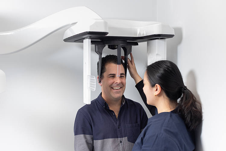
A radiographer will sit you in the centre of the OPG machine and then carefully position and secure your head.
The machine is adjusted for your height and a bite-block is placed between your teeth to ensure correct alignment.
You will need to keep very still during the imaging process, while the rotating arm moves in a semicircle around you, which usually takes around 20 seconds.
An OPG x-ray requires no special preparation and you can return to normal activities straight after your test.
After your appointment
Dental imaging requires no special post treatment care and the patient can return to normal activities straight after the test, as there are no after effects.
One of our radiologists will interpret the scan and provide your referring doctor with a comprehensive report about the findings.
The results of your scan will be sent directly to your referring doctor, and it is very important you book a timely follow-up appointment to discuss your results.
Frequently Asked Questions
No special preparation is required, but patients will be required to remove any jewellery or metallic items from the head and neck region.
At the time of the procedure on the day, our healthcare team will discuss the detail of what will happen during the procedure.
Prior to your scan and to ensure optimal imaging, you may be asked to change into a gown. A change cubicle will be provided to ensure your privacy and you will be asked to bring your belongings will you, carry baskets are provided. You will be asked to place your belongings in a suitable location within the room for the duration of your scan. After your scan, you will be provided with a change cubicle to ensure your privacy. Please ensure you have all your belongings with you prior to leaving the department. If you accidentally leave anything behind, please contact our staff to advise and we will endeavour to locate your belongings and return them to you.
A Dental examination can take from 20 – 30 minutes to complete, depending on the examination requested.
The results and images will be sent to your referring doctor.
Our friendly reception staff will advise you on the cost of your procedure at the time of your booking. If your exam is Medicare eligible, you will be bulk billed.
Patient Safety
Dental scans involve a very small dose of radiation, which makes this test unsuitable for women who are, or might be pregnant.
The benefits of dental scans out outweigh the potential risks involved.