
Our services
Breast Imaging
Our team is made up of highly trained and caring staff who share a dedicated commitment to trusted breast care. We offer comprehensive services including breast screening and diagnostics in a welcoming environment specifically tailored for you. We perform both screening and diagnostic mammograms to ensure your best health.
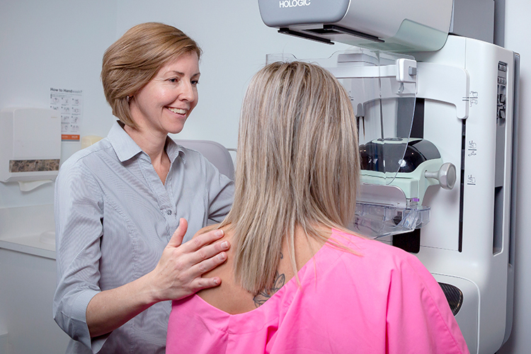
What is Breast Imaging?
At Apex Radiology, your well-being is our top priority. We are committed to using the latest advancements in technology and techniques to provide you with the highest standard of care.
Our multi-modality approach to breast imaging ensures that we can compare and contrast findings from different imaging techniques, enhancing diagnostic accuracy and improving patient outcomes. By employing a combination of these imaging methods, our radiologists can better evaluate and understand breast health, leading to early detection and prompt treatment if necessary.
Types of Breast Imaging Scans
At Apex Radiology, we provide a wide range of breast imaging services to ensure accurate comparison between modalities ensuring the best patient outcomes.
Each type of imaging has its own particular use and is often complementary to other types of imaging.
Our breast imaging services include:
The quality of images produced by MRI of breast tissue is outstanding. It shows much greater detail compared to ultrasound or x-ray mammography.
This gives radiologists the flexibility to examine and view your breast tissue in detail, from many different angles without you needing to change positions. In comparison, mammography requires the breast and the x-ray equipment to be adjusted for each view.
It also makes it easy to take additional images of the muscle and chest wall around your breast to provide a more comprehensive diagnosis.
Breast MRI can be beneficial in detecting breast cancer in high-risk women with a strong family history or past history of breast cancer, or who carrying a genetic predisposition to breast cancer. Therefore, this service may also be used to examine the extent of breast cancer after a diagnosis has been made by mammography or ultrasound and tissue biopsy.
For examining breast implants, mammography and ultrasound may not penetrate saline or silicone well enough to view the implants or surrounding breast tissue. In contrast, MRI may offer clearer images of both implants and breast tissue. This makes MRI an excellent method to assess breast implants and detect small ruptures or leaks.
Before Your Scan
A referral from your doctor and an appointment is required for a breast MRI scan.
This exam is to be booked days 7-11 of your cycle (if you still have cycles).
If you have irregular cycles, a “Serum Progesterone Test” may be organised by Apex Radiology staff.
As soon as the test has been taken, please ensure to call our bookings team to let us know so we can have our specialist Breast Radiologist review your results of this test.
Please arrive 30 minutes prior to your appointment time.
During Your Scan
When you attend your appointment you will be asked to answer a series of questions, remove any jewellery and change into an examination gown. Then the scanning procedure will be explained to you.
The scans are taken as you lay on your stomach. This ensures your breasts are correctly positioned and gently compressed. It will take 30 to 40 minutes to complete the assessment.
For most scans, an injection of the safe contrast solution, gadolinium, into your arm vein will be required—to enhance the detail of the images produced.
After preliminary scans are taken, the contrast solution is injected and the images are acquired.
For scans looking for ruptures to breast implants, these ruptures show up clearly with MRI and the contrast solution is not required.
After Your Scan
After the examination you should be able to resume your normal activities immediately.
What are the risks and side effects?
There are no known side effects.
Who will perform my breast MRI and write the report?
How do I obtain my results?
If the results of your scan are required urgently or you have an appointment with your doctor straight after the examination, please let the Apex Radiology staff know in advance. Otherwise the results of your scan will be sent directly to your referring doctor typically within 48 hours.
Please ensure that you make a follow up appointment with your referring doctor or health care provider to discuss your MRI results.
A Vacuum-assisted stereotactic core biopsy (VAB) is performed to help determine the nature or diagnosis of a breast abnormality and to plan treatment if necessary.
A sample of breast tissue is removed under mammographic guidance for examination by a pathologist, allowing the area for biopsy to be accurately identified, using a special needle with suction to help get the sample from the breast.
Before Your Scan
A referral from your doctor or medical specialist and an appointment is required for this examination.
Please let us know when making your appointment if you are taking medication that makes you bruise or bleed easily.
During Your Scan
When you arrive at Apex Radiology you will be asked to remove all jewellery and clothing from the waist up, and change into a loose fitting examination gown.
VAB is performed while sitting or lying in a special chair with the breast compressed in the same way as for a mammogram.
Several keyhole images will be taken to accurately identify the area for the biopsy. The skin of the breast is then washed with antiseptic before a very fine needle is used to give local anaesthetic to numb the breast in the area for biopsy. The local anaesthetic may sting for a few seconds when it is being given, and then the area will become numb. A small incision will be made and several samples taken. The biopsy procedure may sometimes feel uncomfortable but is usually not painful because of the local anaesthetic that has been given. The process of taking the biopsy lasts just a few minutes.
After the samples have been taken, the biopsy area will be held firmly for a few minutes to minimise any bruising or bleeding, and then covered with a dressing that will be checked before you leave.
Please allow about an hour for the procedure.
After Your Scan
After the procedure, the biopsy site may be tender or show some bruising.
You may place an ice pack over the biopsy site for 20 minutes.
We suggest taking paracetamol to reduce any discomfort.
You will be able to leave the clinic shortly after the procedure.
- You may drive yourself home after the procedure; alternatively, you may prefer to bring someone with you for support and to drive you home.
- Most people find they can return to work the same day.
- You should refrain from exercise for 24 hours following your biopsy.
- Your dressing may be worn in the shower and removed after two to three days.
- If your breast becomes red, swollen or tender in the days after your core biopsy please consult your referring doctor or contact our clinic to check the biopsy site.
Who will perform my vacuum-assisted stereotactic core biopsy and write the report?
At Apex Radiology your Vacuum-assisted stereotactic core biopsy will be performed by a radiologist (a doctor specialising in medical imaging), who will prepare and send a report to your referring doctor detailing the procedure that has been performed.
The results from the core biopsy will be provided by the Pathology provider that the sample is sent to.
How do I obtain my results?
Results from VAB are usually available within 48 hours, but may take up to a week.
Our nurses will ensure that you have an appointment either in our results clinic or with your referring doctor to get your results as soon as possible.
A Breast core biopsy is a quick and simple procedure to perform, which removes a sample of tissue from the breast to be examined by a pathologist under a microscope.
A core biopsy is performed to help determine the nature or diagnosis of a breast abnormality and to plan treatment if necessary.
At Apex Radiology, the breast core biopsy is performed under ultrasound guidance, this shows an image of the inside of your breast on a screen to allow the doctor to visualise the needle is correctly placed.
Before Your Scan
A referral from your doctor or medical specialist and an appointment is required for this examination.
Please let us know when making your appointment if you are taking medication that makes you bruise or bleed easily.
During Your Scan
When you arrive at Apex Radiology you will be asked to remove all jewellery and clothing from the waist up and change into a loose fitting examination gown.
The skin of the breast is then washed with antiseptic and a very fine needle is used to give local anaesthetic to numb the breast in the area for biopsy.
The local anaesthetic may sting for a few seconds when it is being given and after this the area numb. A small nick or cut is made in the skin and the biopsy needle is gently inserted into the breast using the ultrasound for guidance.
Several samples will be taken. When each sample is taken there is a clicking noise, and you may have a feeling of pressure in the breast where the sample is taken.
The biopsy procedure may sometimes feel uncomfortable but is not usually painful because of the local anaesthetic that has been given.
Taking the biopsy samples takes just a few minutes.
After the samples have been taken, the biopsy area will be held firmly for a few minutes to minimize any bruising or bleeding, and then covered with a dressing.
Please allow about an hour for the procedure.
After Your Scan
After your breast biopsy the biopsy site may be tender or show some bruising.
You may place an ice pack over the biopsy site for no more than 20 minutes.
We suggest taking paracetamol for discomfort.
You will be able to leave the clinic shortly after the procedure.
- You may drive yourself home after the procedure.
- Most people can return to work the same day.
- You should refrain from exercise for 24 hours following your biopsy.
- Your dressing may be worn in the shower and removed after two to three days.
- If your breast becomes red, swollen or tender in the days after your biopsy please consult your doctor or contact the Apex Radiology clinic to review the biopsy site.
Risks & Side Effects
Breast core biopsy is a safe procedure, but it is not uncommon for some bruising to appear on the skin at the biopsy site over the next few days.
Sometimes the bruise may develop within the breast tissue causing a tender lump that may take one to two weeks to disappear.
Infection is rare following a core biopsy; please let your doctor know if you develop symptoms after your biopsy.
Who will perform my ultrasound guided core biopsy and write the report?
At Apex Radiology your core biopsy will be performed by a radiologist (a doctor specialising in medical imaging), who will send a report to your referring doctor detailing the procedure that has been performed.
The results from the core biopsy will be provided directly to your referring doctor by the Pathology provider that the sample is sent to.
How do I obtain my results?
The results from the core biopsy will be provided directly to your referring doctor by the Pathology provider that the sample has been sent to.
Results from core biopsy are usually available within 36 hours, but can take up to 5 days.
Please ensure that you make a follow up appointment with your referring doctor or health care provider to discuss your biopsy results.
3D Mammography (Tomosynthesis) is new breast imaging technology that has been proven to improve the accuracy in diagnosis of breast cancer. It is performed as part of a diagnostic mammogram examination.
In addition to the standard mammographic views, a specialised view (tomographic) is taken to produce 3-D images. Tomographic view is completed within seconds from a single sweep of the x-ray arm, creating a series of detailed images that together make up the 3D imaging of the breast.
A 3D Mammography (Tomosynthesis) mammogram may be requested by your referring doctor to gain information about a current breast symptom or as a follow up test to an existing condition.
Mammograms may also be requested if you have a strong family history of breast cancer. Mammography plays an important role in the early detection of breast cancer because it can show changes in the breast long before they can be felt.
Before Your Scan
A referral from your doctor and an appointment is required for this examination.
If you experience tenderness in the breast before your menstrual period, do not schedule the examination during this time.
The best time is one week following your menstrual period, unless the examination is urgent.
If you have breast implants or require wheelchair access, please advise our staff at the time of making your appointment as we may need to allocate a longer appointment time.
During Your Scan
When you arrive at Apex Radiology you will be asked to remove all jewellery and clothing for the waist up and change into a loose fitting examination gown.
Initially two (or sometimes more) images will be taken of each breast.
While the mammogram is being taken, each breast will be gently compressed between two flat plates on the x-ray machine.
For the tomographic view, the x-ray arm will move in an arc above the breast for a short time.
Some women may experience discomfort with compression; however, if you experience pain during the mammogram you should inform the radiographer. You can also ask for the procedure to stop at any time.
We recommend that you advise the radiographer if you have sensitive breasts, they will work with you to make sure that the mammogram is as comfortable as possible.
Unfortunately, compression of the breast is essential to ensure an accurate image and minimise the amount of radiation used.
After the routine views of your breast have been obtained the radiographer will ask you to wait while they are examined by a radiologist to ensure that all the images needed have been obtained.
A breast ultrasound is often requested as a complementary test at the time of the mammogram or at a later date as a means of gathering more information for a complete examination.
After Your Scan
You can resume normal activity immediately after your examination.
As a result of compression required during the examination, you may experience mild tenderness afterward this should subside within 24hrs.
Risks & Side Effects
Your doctor has sent you for a mammogram knowing that the information provided by the scan is more beneficial than the very low radiation risk associated with it.
The radiation dose using tomosynthesis is only slightly higher than standard mammography and is well within the Australian National Guidelines for radiation safety levels.
The skill of the operator and the design of the machine ensure you receive the lowest radiation dose possible.
Female patients who are pregnant or think they may be pregnant must advise staff prior to the examination, as a mammogram is not usually performed during pregnancy unless it is a medical necessity.
Who will perform my mammogram and write the report?
Your mammogram will be carried out by a radiographer with specific training in mammography, they have a degree in medical imaging and are accredited by the Australian Institute of Radiography (AIR).
Your images will be examined by a Apex Radiology radiologist (a medical doctor specialising in the interpretation of medical images), accredited by the Royal Australian and New Zealand College of Radiologists, who will prepare and send a report to your referring doctor or medical specialist detailing the findings of your examination.
How do I obtain my results?
If your mammography results are considered to be medically urgent, or you have an appointment with your referring doctor or health care provider straight after your examination, Apex Radiology will arrange to have your results available immediately. Otherwise your referring doctor or health care provider will receive your report within 48 hours of your examination.
Please ensure that you make a follow up appointment with your referring doctor or health care provider to discuss your results.
A Breast Fine Needle Aspiration (Ultrasound Guided Fine Needle Aspiration) (FNA) is a quick and simple procedure to remove fluid or cells from a breast lesion or cyst (a lump, sore or swelling) with a fine needle similar to a blood sample needle.
The sample fluid or cells are sent to a pathology laboratory to be examined by a specialist doctor (a cytologist).
An FNA is performed to help determine the nature or diagnosis of the lesion and to plan treatment if necessary.
Breast FNA may also be used to aspirate a cyst using the needle draw fluid from the cyst to remove it completely or relieve discomfort if the cyst is large and tender.
Before Your Scan
A referral from your doctor or medical specialist and an appointment is required for this examination.
Please let us know when making your appointment if you are taking medication that makes you bruise or bleed easily.
During Your Scan
When you arrive at Apex Radiology you will be asked to remove all jewellery and clothing from the waist up, and change into a loose fitting examination gown.
The procedure will be explained and you will be asked to lie on the examination table.
The skin of your breast washed with antiseptic.
A thin needle (similar to a needle used for taking a blood sample), is placed through the skin into the breast, often using ultrasound guidance, to sample the area of interest.
The needle stays in the breast for a short time, while it is gently moved back and forth to enable cells to be collected. Please allow about an hour for the procedure.
After Your Scan
After your Breast FNA procedure, the biopsy site may be tender or show some bruising.
You may place an ice pack over the site for no more than 20 minutes.
To relieve any discomfort we suggest taking paracetamol; otherwise, no special care is required.
- You will be able to leave the clinic immediately after the procedure.
- You may drive yourself home after the procedure.
- You may resume your normal activities.
- You may remove the adhesive dressing on the evening of the FNA
- If your breast becomes red, swollen or tender in the days after your FNA, please consult your referring doctor or contact the Apex Radiology to review the Breast FNA site.
Risks & Side Effects
Breast FNA is a safe procedure, it is common to get a small bruise on the skin following an FNA and occasionally a bruise may develop in the breast tissue which causes a tender lump. This usually settles within a few days. Infection is rare following FNA. Please let your doctor know if you develop any concerning symptoms after the FNA.
Who will perform my ultrasound guided FNA and write the report?
At Apex Radiology your Breast FNA will be performed by a radiologist (a doctor specialising in medical imaging), who will send a report to your referring doctor detailing the procedure that has been performed.
The results from the FNA will be provided directly to your referring doctor by the Pathology provider that the sample is sent to.
How do I obtain my results?
The results from the Breast FNA will be provided directly to your referring doctor by the Pathology provider that the sample has been sent to. Results from FNA are usually available within 36 hours but can take up to 5 days. Sometimes results from FNA may be inconclusive and require repeat or further tests.
Please ensure that you make a follow up appointment with your referring doctor or health care provider to discuss your results.
Mammography, ultrasound and magnetic resonance imaging (MRI) examinations can identify abnormalities in the breast that cannot be felt by a doctor.
If the abnormality is to be surgically removed, it is necessary to place a fine wire (called a hookwire), into the breast with its tip at the site of the abnormality.
The wire acts as a marker during surgery and enables the surgeon to identify the correct area of breast tissue.
Mammography, ultrasound or MRI scans are used to guide the hookwire into the correct position.
The wire is called a hookwire because there is a tiny hook at the end, which keeps it in position.
Before Your Scan
A referral from your doctor and an appointment are required for this procedure.
Usually, this procedure will be performed a few hours before you have surgery.
There is no preparation required for the hookwire localisation, but there will be preparation for the surgery that follows the hookwire localisation.
Preparation instructions/information for the surgery will provided to you by your surgeon.
During Your Scan
Before the procedure you will be asked to remove all jewellery and clothing from the waist up, and change into a loose fitting examination gown.
The skin of the breast will then be washed with antiseptic before a very fine needle is used to give local anaesthetic to numb the breast in the area for biopsy.
The local anaesthetic may sting for a few seconds when it is being given, and after this the area will become numb.
The radiologist will then insert a fine needle into the tissue to be removed.
Images will be taken to check the position of the needle, once it is in the correct position, a fine wire is passed through the centre of the needle and the needle is removed, leaving the hookwire in place.
A final set of images will be taken to show the surgeon where the tip of the wire lies in relation to the abnormality that is to be removed.
After Your Scan
Following the hookwire placement, a piece of the fine wire will be protruding from the breast.
This projecting wire will be taped down to the skin and the hookwire remains in the abnormality in the breast.
The surgeon will remove the wire together with the abnormality at the time of the operation.
Your previous imaging and the images from the Hookwire Localisation will be sent with you to the operating theatre so that the surgeon may refer to them.
Risks & Side Effects
Hookwire localisation is a simple procedure to perform and most women will experience no problems.
Problems that can occur on rare occasions are:
- Movement of the hookwire after placement and before surgery is performed (which reduces the accuracy of the surgery), and
- Wire dislodgement. This occurs usually because the breast is composed of fatty tissue which provides a poor grip for the hookwire).
If you are travelling to another facility for your surgery with a hookwire in position, you need to take care. Dislodgement may occasionally occur with very little movement. If dislodgement occurs, you may need to have the procedure repeated because the tip of the wire will no longer be situated in the lesion that needs to be removed.
Who will perform my hookwire localisation and write the report?
At Apex Radiology your hookwire localisation procedure will be performed by a radiologist (a doctor specialising in medical imaging), who will send a report to your surgeon detailing the procedure that has been performed.
If you have noticed a change in your breasts or a mammogram has detected an abnormality, your general practitioner may request you to have a breast ultrasound.
A breast ultrasound is a non-invasive, painless test that uses sound waves to form an image of structures in the breast, which helps your healthcare provider determine whether these changes are caused by a non-cancerous or benign lump of tissue or a breast cancer based upon the shape, location and other characteristics visible on ultrasound.
Before Your Scan
You will need a referral from your doctor to make an appointment.
Read all the instructions given to you by your doctor or provided by us, as you may be required to fast depending on the type of imaging.
Wear clothing that will provide easy access to the area requiring imaging.
Please bring any previous, relevant examinations for comparison.
If you have diabetes, or you are on any medication, please contact us to check special preparation instructions, and our staff will advise of any specific preparation.
During Your Scan
The sonographer, the medical professional administering your breast ultrasound, will spread gel on the area/s to be scanned.
They will then move a small device called a transducer over your breast and the lymph nodes in your armpit. The scan usually takes 15-20 minutes.
After Your Scan
After your scan you can go about your day as normal.
One of our radiologists will interpret the ultrasound and provide your referring doctor with a comprehensive report about the findings.
It is very important you book a timely follow-up appointment to discuss your results.
About Your Test
Before your appointment
You will need a referral from your doctor to make an appointment.
Upon receiving your referral, our Bookings Team will be able to help assist you in finding a time that works for you to have your test done.
On the day
After your appointment
After your scan you can go about your day as normal.
Your images will be looked over by our Radiologists and a secure report and copy of the images will be provided to your referring practitioner. We will also provide you with a printed copy to take with you to your next appointment.
Frequently Asked Questions
One of our Mammographers (specially trained radiographers) will explain and guide you through the procedure. They will ask about your relevant medical history which may include information about any current symptoms and your family history. The mammographer will gently position each breast, one at a time for each picture. Your breast is compressed with a plastic plate for just a few seconds. Using compression is very important to allow us to get clear and detailed images. Generally, at least 2 views of each breast will be taken. You are welcome to ask any questions and encouraged to discuss with your Mammographer if you experience any discomfort.
For both screening and diagnostic mammograms we utilise Digital Breast Tomosynthesis or 3D Mammography. During your mammogram the Tomosynthesis machine sweeps in a small arc above your breast. This produces images in layers as thin as one millimetre. We can now view your breast images one slice at a time, like turning the pages of a book.
Two fully accredited Breast Diagnostic Specialists will independently review your images. We will send a report of your results to your referring Doctor (GP or specialist) to discuss with you at your follow-up appointment.
You are welcome to consult with one of our specialists on the day to ask any questions you may have.
We ask that you bring your referral and any previous mammogram films or breast imaging that you have had. This is important so we can perform a detailed comparison report. You also need to bring your Medicare or Pension Card. A two-piece outfit may be more convenient on the day when you are changing.
Your radiation exposure is within the Australian Radiation Protection and Nuclear Safety Agency (ARPANSA) guidelines.
Your images and report are provided through a secure online system to your doctor or healthcare provider. We will also provide you with a printed copy to bring to your next appointment.
Our friendly reception staff will advise you on the cost of your procedure at the time of your booking. If your exam is Medicare eligible, you will be bulk billed.
Prior to your scan and to ensure optimal imaging, you may be asked to change into a gown. A change cubicle will be provided to ensure your privacy and you will be asked to bring your belongings will you, carry baskets are provided. You will be asked to place your belongings in a suitable location within the room for the duration of your scan. After your scan, you will be provided with a change cubicle to ensure your privacy. Please ensure you have all your belongings with you prior to leaving the department. If you accidentally leave anything behind, please contact our staff to advise and we will endeavour to locate your belongings and return them to you.
Breast Screening Mammograms are performed when you have no symptoms as part of a proactive approach to breast health.
Diagnostic Mammograms are performed if you have abnormalities or symptoms your Doctor asks to be investigated.
We perform both screening and diagnostic mammograms.