
Our services
Pregnancy Ultrasound
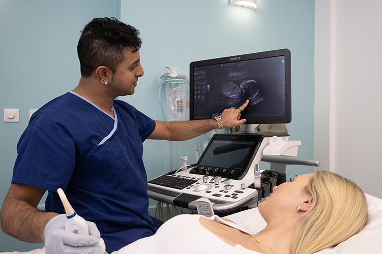
Pregnancy ultrasound is a non-invasive painless examination to assess fetal growth, position and anatomy, and placental site, as well as the maternal uterus and ovaries.
The ultrasound produces a real time moving image on a screen.
What is Pregnancy Ultrasound?
Ultrasound provides useful information about the uterus, ovaries and baby that enables appropriate antenatal care.
Fetal size and heart rate are checked at all stages of pregnancy, as well as the maternal uterus, ovaries and cervix.
Pregnancy ultrasound is a non-invasive painless examination to assess fetal growth, position and anatomy, and placental site, as well as the maternal uterus and ovaries.
Ultrasound can detect many, but not all abnormalities. Demonstrating a fetal abnormality on ultrasound depends on many factors, including fetal age and position at the time of the ultrasound, and the size and type of abnormality.
Types of Pregnancy Ultrasound
During your pregnancy you will have a series of ultrasounds to assess your baby’s development.
This examination may be performed internally, externally or both. The best way to examine the pelvic organs in detail is to perform a transvaginal (endovaginal) ultrasound, in which the ultrasound transducer is on the end of a thin probe which is inserted into the vagina.
Transvaginal ultrasound is usually recommended for patients who are 18 years and above. If the examination is not urgent, it is best performed between days 5 to 12 of your menstrual cycle. The sonographer will explain the process in detail and ensure that you are happy to have the examination – it will not be performed without your consent.
External pelvic ultrasound – In situations where an internal pelvic ultrasound is not appropriate, the examination will be performed by placing the ultrasound transducer on top of the lower abdomen.
To ensure that the inside of the pelvis area is seen clearly on the screen, a full bladder is required. Empty your bladder one (1) hour before your appointment, then drink one (1) litre of clear fluid or cordial. Do not empty your bladder before your appointment.
During the second trimester, a Morphology Scan (18-22 weeks) is used to check fetal growth, numerous important organs such as the spine, heart, brain and kidneys, placental site, and amniotic fluid volume.
During the morphology scan, the sonographer will check for signs of a possible genetic problem.
All Australian women are encouraged to have a morphology scan, and Medicare will provide a rebate for one or more scans during this period depending on the clinical situation and whether an obstetrician or GP refers the patient.
During the third trimester, an ultrasound can check the fetal position, placental appearance and site, how much fluid is around the baby, fetal well-being and the baby’s growth compared with previous scans and the expected date of delivery.
About Your Test
Before your appointment
You will need a referral from your doctor to make an appointment.
Wear clothing that will provide easy access to the area requiring imaging.
Please bring any previous, relevant examinations for comparison.
If you have diabetes, or you are on any medication, please contact us to check special preparation instructions, and our staff will advise of any specific preparation.
On the day
During the examination a water based gel will be applied to the skin over the area to be scanned to improve the transmission of sound waves between the body and the ultrasound transducer.
To get the best possible images the sonographer may ask you to hold your breath or move your position so the best images can be obtained.
On occasion, a radiologist may need to speak with you and perform some ultrasound scanning as well, in order to clarify what has been shown in the images and ensure the question asked by your referring doctor is answered accurately.
After your appointment
After your scan you can go about your day as normal.
One of our radiologists will interpret the ultrasound and provide your referring doctor with a comprehensive report about the findings.
It is very important you book a timely follow-up appointment to discuss your results.
Frequently Asked Questions
Please fill your bladder based on the information our reception staff will provide at the time of booking. Also please bring in any prior scans you may have for this pregnancy.
Prior to your scan and to ensure optimal imaging, you may be asked to change into a gown. A change cubicle will be provided to ensure your privacy and you will be asked to bring your belongings will you, carry baskets are provided. You will be asked to place your belongings in a suitable location within the room for the duration of your scan. After your scan, you will be provided with a change cubicle to ensure your privacy. Please ensure you have all your belongings with you prior to leaving the department. If you accidentally leave anything behind, please contact our staff to advise and we will endeavour to locate your belongings and return them to you.
Your images and report are provided through a secure online system to your doctor or healthcare provider. We will also provide you with a printed copy to bring to your next appointment.
Our friendly reception staff will advise you on the cost of your procedure at the time of your booking. If your exam is Medicare eligible, you will be bulk billed.
Patient Safety
Ultrasound is a safe examination which provides excellent imaging without any known risks.
Our staff use ultrasound with care to ensure that patients benefit from what ultrasound offers, with minimum risk.
At Apex Radiology we employ experienced, qualified sonographers accredited with the Australian Sonographer Accreditation Registry, and invest in the latest equipment to ensure patients receive the very best patient care.